
Anti-Prostasin Rabbit Polyclonal Antibody

- Host
- Rabbit, Polyclonal
- Reactivity
- Validated- Human Potential-Mouse, Rat, Pan, Monkey, Dog
- UniProt
- Q16651
- Size
- 100ug
- Cat. #
- RP2Prostasin
In stock
- SKU
- RP-Prostasin
Target Overview
Prostasin (PRSS8, UniProt Q16651) is a 343-amino acid, GPI-anchored serine protease of the trypsin family (EC 3.4.21.-) localized to the cell membrane. Also known as channel-activating protease 1 (CAP1), prostasin exhibits trypsin-like substrate specificity with a preference for poly-basic cleavage sites. The protease is synthesized as a single-chain zymogen and processed into disulfide-linked light and heavy chains, with the heavy chain containing the catalytic triad. Prostasin plays a critical role in epithelial sodium transport by proteolytically activating the epithelial sodium channel (ENaC) through cleavage of the gamma subunit (SCNN1G). Originally identified in seminal fluid and prostate, prostasin is broadly expressed in epithelial tissues including kidney, lung, colon, and skin. Researchers study prostasin in the context of epithelial ion homeostasis, blood pressure regulation, and barrier function. Recent work has identified prostasin as an endogenous sheddase for TGFBR3 and highlighted its appearance in plasma proteomic signatures associated with hypertension, inflammatory bowel disease, and psoriatic conditions.
Background
References
- Greulich BM et al (2026) Human PRSS8 functions as a targetable, endogenous TGFBR3 sheddase via the novel cholesterol-PRSS8-TGF-β axis. Biochem Biophys Res Commun. PubMed · doi:10.1016/j.bbrc.2026.154011
- Zhang H et al (2026) Plasma proteomic profiles reveal proteins and characteristic patterns associated with hypertension: A prospective cohort study. J Hypertens. PubMed · doi:10.1097/HJH.0000000000004327
- Abebe E et al (2026) Uncovering plasma protein biomarkers linked to depression: A differential abundance analysis and Mendelian randomization using large-scale data. J Affect Disord. PubMed · doi:10.1016/j.jad.2025.121134
- Feng J et al (2025) Plasma proteomic profiles identify biomarkers predicting Crohn's disease up to 16 years before onset. Nat Commun. PubMed · doi:10.1038/s41467-025-66483-4
- Wu T et al (2026) Population-Based Multi-Omics and Cohort Study Identifying Predictive Biomarkers and Therapeutic Targets for Psoriatic Disease. Adv Sci (Weinh). PubMed · doi:10.1002/advs.202514130
Additional Specifications
| Gene Symbol | PRSS8 |
|---|---|
| UniProt ID | Q16651 |
| Host Species | Rabbit |
| Species Reactivity | Validated- Human Potential-Mouse, Rat, Pan, Monkey, Dog |
| Pack Size | 100ug |
| Immunogen (amino end of the catalytic domain) | Immunogen is proprietary and confidential. Immunogen generated in amino acid region 45-85. |
| Immunogen (Catalytic domain) | Immunogen is proprietary and confidential. Immunogen generated in amino acid region 45-286. |
| Immunogen (carboxy end of the catalytic domain) | Immunogen is proprietary and confidential. Immunogen generated in amino acid region 246-286. |
| Immunogen (Linker Region) | Immunogen is proprietary and confidential. Immunogen generated in amino acid region 290-318. |
| Alternate Names | PRSS8, Channel-activating protease 1, CAP1, Serine protease 8, Prostasin light chain, Prostasin heavy chain |
Frequently Asked Questions
What molecular weight band should I expect for prostasin on a Western blot?
Prostasin is synthesized as a single-chain 343-amino acid zymogen with a predicted mass of approximately 40 kDa. Following post-translational processing, the mature form is cleaved into disulfide-linked light and heavy chains. On reducing SDS-PAGE, you may observe bands around 28–30 kDa (heavy chain containing the catalytic domain) and 10–12 kDa (light chain). Under non-reducing conditions, expect the mature heterodimer near 40 kDa. Glycosylation can introduce minor shifts. The GPI anchor is cleaved during sample preparation, so you are detecting the solubilized protease forms, not the membrane-tethered species.
What is the recommended starting dilution for prostasin antibody in Western blot?
We recommend starting at 1:1000 dilution for Western blot, based on validation with human lysates. This dilution typically yields clean signal with low background when using standard chemiluminescent detection. If your tissue or cell line expresses prostasin at lower levels—common in non-epithelial samples—consider concentrating your lysate or increasing antibody concentration to 1:500. Conversely, kidney cortex and certain epithelial lines express prostasin abundantly; you may achieve good signal at 1:2000. Always include a positive control lysate from prostate or renal epithelial cells to confirm antibody performance in each experiment.
Does this prostasin antibody cross-react with mouse and rat samples?
The antibody is validated for human prostasin. Cross-reactivity with mouse and rat is predicted based on sequence homology—human and mouse prostasin share approximately 70 percent identity in the immunogen region—but has not been experimentally validated by Triple Point Biologics. Researchers have reported successful detection in mouse kidney and lung lysates at similar dilutions, though band intensity may differ. If working with rodent samples, include a known positive control and verify band size. Non-human primate, canine, and porcine sequences show strong homology and are also predicted to cross-react, but empirical testing in your system is essential.
What sample preparation considerations are important for detecting membrane-anchored prostasin?
Prostasin is GPI-anchored at the plasma membrane, so complete solubilization requires detergent-based lysis. RIPA buffer or buffers containing 1 percent Triton X-100 or NP-40 work well. Avoid detergent-free lysis, which will partition prostasin into the insoluble membrane fraction. Prostasin is also shed into extracellular fluid as a soluble protease following GPI cleavage by endogenous phospholipases; conditioned media from epithelial cultures can be concentrated and blotted directly. For tissue samples, enrich for epithelial compartments when possible. Snap-freeze samples immediately, as prostasin is an active protease and may undergo autolysis or degrade other proteins during prolonged handling.
Which positive and negative control cell lines should I use for prostasin Western blots?
Positive controls include LNCaP or PC-3 prostate carcinoma cells, which express robust prostasin, and primary renal proximal tubule or cortical collecting duct cells. Human bronchial epithelial lines (e.g., 16HBE) also express detectable prostasin. For tissue lysates, kidney cortex and seminal fluid are strong positives. Negative controls are less straightforward—prostasin is broadly expressed in epithelia at varying levels. Mesenchymal cell lines such as NIH/3T3 fibroblasts or HEK293 cells show low to absent expression and serve as useful negative or low-expression comparators. Verify each lot by running your chosen controls in parallel with experimental samples.
Can this antibody distinguish between zymogen and active prostasin forms?
This polyclonal antibody recognizes both the single-chain zymogen and the cleaved, disulfide-linked mature heterodimer, as the immunogen spans residues present in both forms. It does not selectively detect the active site or activation cleavage neo-epitope, so you cannot distinguish zymogen from active protease by immunoreactivity alone. To assess activation state, run samples under reducing versus non-reducing conditions and examine the banding pattern: single-chain zymogen migrates near 40 kDa; the mature form resolves into heavy and light chains under reduction. Alternatively, combine Western blotting with activity assays or use activation-state-specific antibodies if cleavage status is critical to your question.
How should I store the prostasin antibody and what is its shelf life?
Store the antibody at –20°C as supplied. For routine use, prepare small aliquots (e.g., 10–20 µL) to avoid repeated freeze-thaw cycles, which can reduce titer and increase aggregation. An aliquot in working solution can be held at 4°C with 0.02 percent sodium azide for up to one month if you use it frequently. Do not store diluted antibody in blocking buffer for extended periods. Under these conditions, the antibody remains stable for at least two years from the date of receipt. If you observe increased background or diminished signal over time, prepare a fresh aliquot and compare side by side before troubleshooting other variables.
What is the biological relevance of prostasin in epithelial sodium channel regulation?
Prostasin proteolytically activates the epithelial sodium channel (ENaC) by cleaving the extracellular loop of the gamma subunit (SCNN1G), releasing an inhibitory peptide and increasing channel open probability. This mechanism is critical for sodium reabsorption in the kidney collecting duct and for maintaining airway surface liquid volume in the lung. Loss of prostasin activity impairs ENaC-mediated sodium transport, as seen in conditional knockout models exhibiting salt wasting or altered blood pressure. Prostasin itself is regulated by serine protease inhibitors, notably HAI-1 and HAI-2, forming a protease-inhibitor axis that fine-tunes epithelial sodium homeostasis. Dysregulation is implicated in hypertension, polycystic kidney disease, and cystic fibrosis pathophysiology.
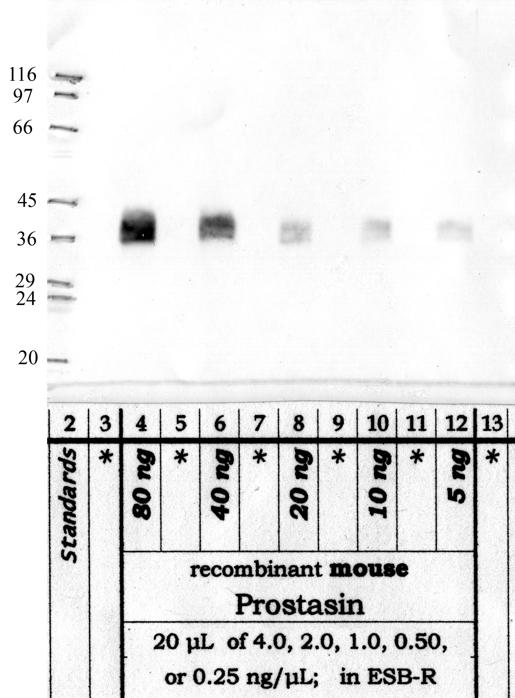
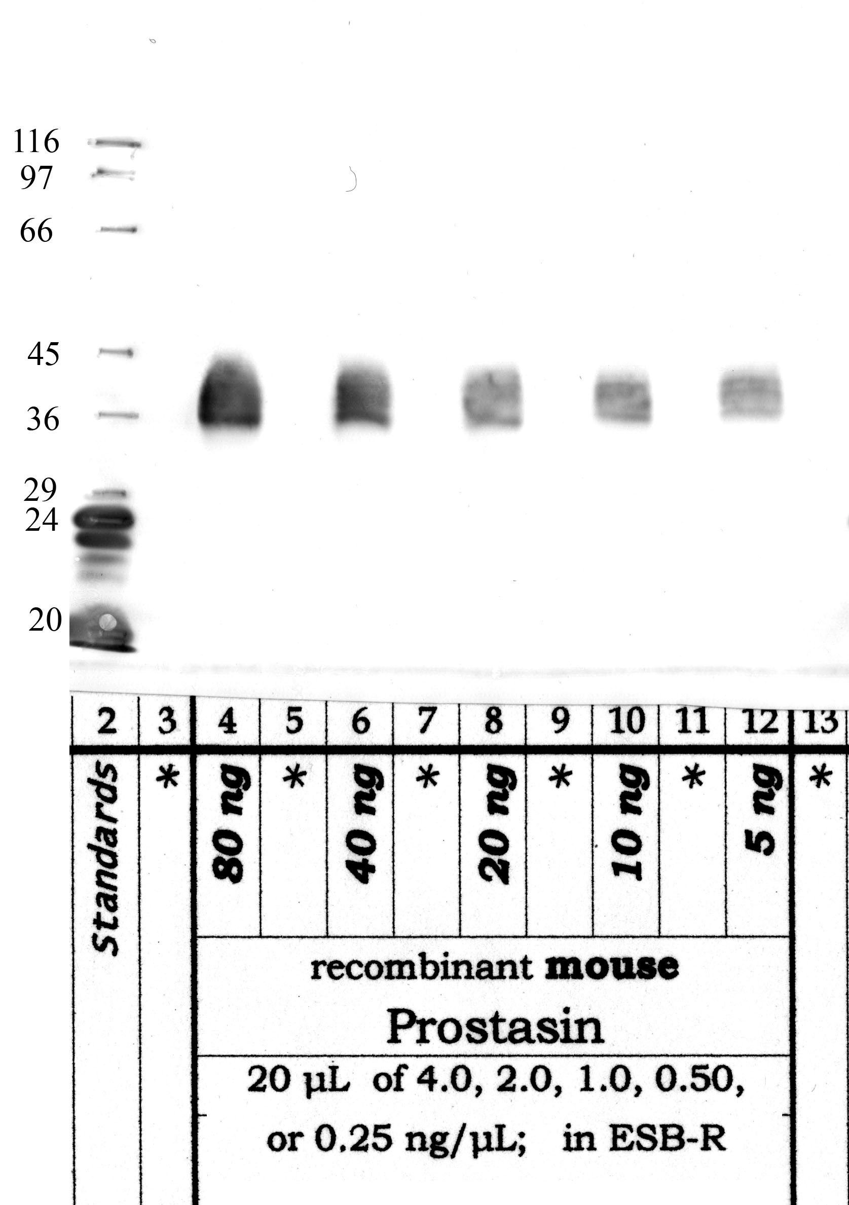
Western blot validation for RP-Prostasin — 4 panels across the domain-specific antibody variants. Each blot below shows the clone that validates a specific domain of the target protein.


Custom validation studies available on request — contact us.
Also known as:
- PRSS8
- Channel-activating protease 1
- CAP1
- Serine protease 8
- Prostasin light chain
- Prostasin heavy chain