
Anti-Furin Rabbit Polyclonal Antibody
- Host
- Rabbit, Polyclonal
- Reactivity
- Validated- Human Potential-Mouse, Rat, Pan, Monkey, Dog
- UniProt
- P09958
- Size
- 100ug
- Cat. #
- RP1Furin
In stock
- SKU
- RP-Furin
Target Overview
Furin (UniProt P09958; EC 3.4.21.75) is a calcium-dependent serine endoprotease and the prototypical member of the proprotein convertase (PCSK) family. The 794-residue type I transmembrane protein cycles between the trans-Golgi network, the cell surface, and endosomes, and is also referred to as PACE (paired basic amino acid residue-cleaving enzyme) or dibasic-processing enzyme. Furin cleaves a broad array of precursor proteins at the consensus motif R-X-(K/R)-R within the constitutive secretory pathway, generating mature, biologically active forms of growth factors, receptors, adhesion molecules, and pathogen-derived proteins. Documented physiological substrates include pro-TGF-β1, pro-BNP (yielding the active BNP(1-32) cardiac hormone), and the V-ATPase accessory subunit ATP6AP1/Ac45, which modulates acidification of dense-core secretory granules. Furin is ubiquitously expressed, with highest activity in liver, kidney, and salivary gland, and its trafficking and activity are tightly regulated by autoinhibitory prodomain cleavage, pH-dependent conformational changes, and cytoplasmic sorting signals. Because furin processes numerous viral envelope glycoproteins—including HIV gp160, influenza HA of highly pathogenic strains, and the SARS-CoV-2 spike at the S1/S2 boundary—it is a longstanding focus of antiviral research. It is also implicated in tumor progression, fibrosis, and cardiovascular remodeling through activation of metalloproteases, IGF receptor, and Notch signaling components. Triple Point Biologics offers two rabbit polyclonal antibodies (RP1Furin, RP2Furin) raised against the amino end of the mature enzyme, validated in human samples by Western blot, with predicted cross-reactivity to mouse, rat, Pan, monkey, and dog based on sequence conservation across the catalytic and P domains.
Background
References
- Seidah NG (2026) The secretory PCSK family in cardiovascular disease and beyond. Atherosclerosis. PubMed · DOI
- Chen S et al (2026) Plasma proteomics reveal key proteins mediating vascular aging. Life Sci. PubMed · DOI
- Santana LADM et al (2026) Alert on a Novel South American Bat Coronavirus with a Furin-Cleavage Site: A One Health Early Warning. Rev Soc Bras Med Trop. PubMed · DOI
- Pan J et al (2026) Fe(3+)-responsive aggregable gold nanoplatform enables siRNA-mediated chemoresistance reversal and combined photothermal-temozolomide therapy for glioblastoma. Acta Biomater. PubMed · DOI
Additional Specifications
| Gene Symbol | FURIN |
|---|---|
| UniProt ID | P09958 |
| Host Species | Rabbit |
| Species Reactivity | Validated- Human Potential-Mouse, Rat, Pan, Monkey, Dog |
| Pack Size | 100ug |
| Immunogen (Amino end mature enzyme) | Immunogen is proprietary and confidential. Immunogen generated in amino acid region 108-158. |
| Immunogen (Homo-B domain) | Immunogen is proprietary and confidential. Immunogen generated in amino acid region 599-672. |
| Alternate Names | Furin, FUR, PACE, Paired basic amino acid residue-cleaving enzyme, Dibasic-processing enzyme, PCSK3, Proprotein convertase subtilisin/kexin type 3, EC 3.4.21.75 |
Frequently Asked Questions
What molecular weight should I expect for Furin on a Western blot?
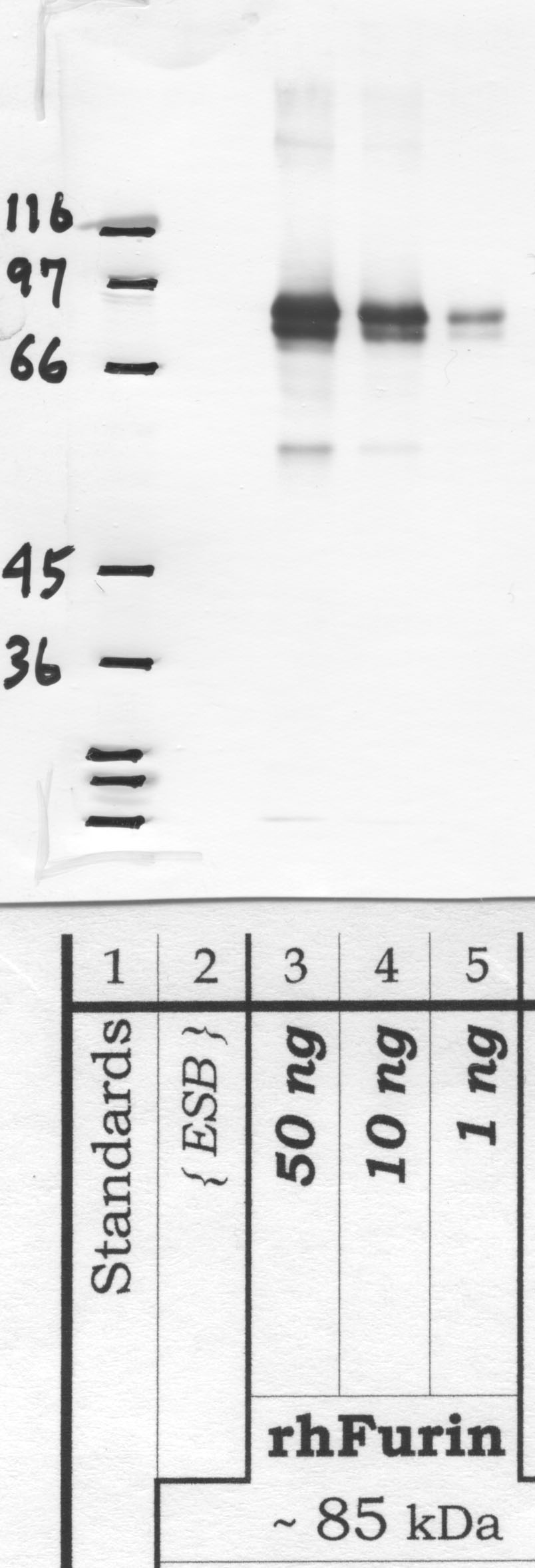
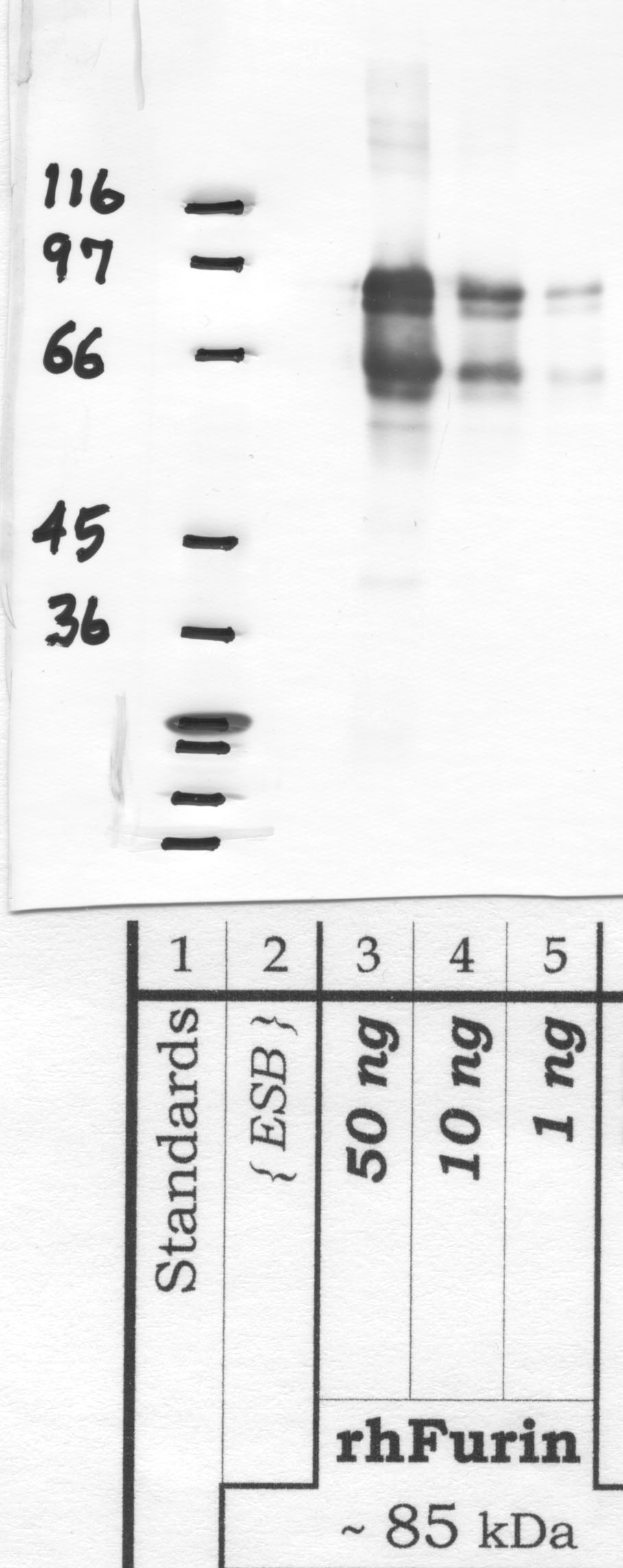
Full-length Furin runs as a ~94 kDa precursor (pro-Furin) and a ~90 kDa mature form following autocatalytic propeptide removal in the ER. Depending on cell type and processing, you may also observe a ~75-80 kDa shed/soluble species lacking the transmembrane and cytoplasmic domains, generated by ectodomain cleavage. Glycosylation can shift apparent mass by several kDa. In overexpression systems, the unprocessed proenzyme is more prominent; in steady-state lysates of secretory cells (HepG2, MDA-MB-231, A549), the mature band typically dominates. Run reducing SDS-PAGE on 8-10% gels for clean resolution.
What positive control lysates work well for this Furin antibody?
HepG2, A549, MDA-MB-231, HeLa, and HEK293 all express endogenous Furin at detectable levels by Western blot. HepG2 is a reliable benchmark because Furin is constitutively active in hepatocyte-derived lines for processing albumin and clotting factor precursors. For a stronger positive control, transient transfection of Furin cDNA into HEK293 or CHO-derived FD11 (Furin-deficient) cells gives a clear gain-of-signal comparison. Loss-of-signal controls using FD11 cells or Furin siRNA knockdown in HepG2 are useful for confirming specificity, since the PCSK family shares conserved catalytic domains and cross-reactivity with PC5/6 or PACE4 is a known concern.
What dilution should I start with for Western blot?
Start at 1:1000 in 5% non-fat milk or BSA in TBST for overnight incubation at 4°C. For low-expressing samples or primary tissue, titre down to 1:500; for overexpression lysates, 1:2000-1:5000 is often sufficient. Furin is a relatively low-abundance endoprotease in most cell types, so loading 30-50 µg total protein per lane is recommended. Use HRP-conjugated anti-rabbit secondary at 1:5000-1:10000. If background is high, switch blocking to 5% BSA, since milk phosphoproteins can contribute non-specific bands in the 80-90 kDa range.
Can I use this antibody for immunofluorescence to track Furin trafficking?
The antibody is validated brand-wide for IF. Furin cycles between the TGN, plasma membrane, and endosomes, with steady-state localization predominantly in the TGN due to the cytoplasmic tail acidic cluster and CK2 phosphorylation-dependent retrieval. Co-stain with TGN46 or golgin-97 to confirm Golgi localization, and EEA1 for endosomal pools. Fix with 4% PFA (10 min, RT) and permeabilize with 0.1% Triton X-100 or 0.05% saponin; saponin preserves Golgi morphology better. Methanol fixation can disrupt the Furin epitope depending on the immunogen region, so PFA is the safer starting point.
Does the antibody cross-react with other proprotein convertases like PC5/6 or PACE4?
Reactivity is validated in human and predicted in mouse, rat, pan, monkey, and dog based on sequence conservation across the immunogen region. The PCSK family (Furin, PC1/3, PC2, PC4, PC5/6, PACE4, PC7) shares a conserved subtilisin-like catalytic domain, so any polyclonal raised against catalytic-domain epitopes carries a theoretical cross-reactivity risk. We have not observed PC5/6 or PACE4 signal in standard validation lysates, but if your system overexpresses other PCSKs, confirm specificity with a Furin-knockout or FD11 control. Tissues with high PACE4 expression (pituitary, ovary) warrant extra care.
Will this antibody detect Furin in mouse or rat tissue?
Reactivity in mouse and rat is predicted based on sequence homology — human Furin shares ~96% identity with mouse and rat orthologs across most of the ectodomain — but not formally validated by TPB. Researchers have reported successful detection in mouse liver and kidney lysates with polyclonal anti-human Furin reagents, though you should expect to re-titrate (often 1:500 for rodent tissue) and include a no-primary control. If you publish rodent data, confirming with a Furin-knockout tissue or siRNA knockdown is the cleanest way to establish specificity in your hands.
Why am I seeing multiple bands between 75 and 95 kDa?
Multiple bands in this range are usually genuine Furin species, not non-specific signal. Pro-Furin (~94 kDa), mature membrane-bound Furin (~90 kDa), and shed soluble Furin (~75-80 kDa) commonly co-exist, with relative ratios depending on cell type, confluency, and serum conditions. N-glycosylation at multiple sites adds heterogeneity; PNGase F treatment will collapse the doublet. Proteolytic clipping during lysis is another source — always lyse cold in RIPA or NP-40 buffer with complete protease inhibitors plus 1 mM EDTA (Furin itself is calcium-dependent, so EDTA inhibits autoprocessing artifacts during prep).
How should I store and aliquot the antibody?
Store at -20°C for long-term and 4°C for short-term (up to one month) use. The 100 µg vial ships in PBS with glycerol and sodium azide as a preservative. Aliquot into low-protein-binding tubes (5-10 µL working aliquots) on first thaw to avoid repeated freeze-thaw cycles, which degrade polyclonal performance over time. Avoid frost-free freezers where temperature cycling occurs. Azide inhibits HRP, so do not include the primary antibody dilution directly with HRP conjugates without a wash step. Centrifuge briefly before pipetting to recover any antibody from the cap.
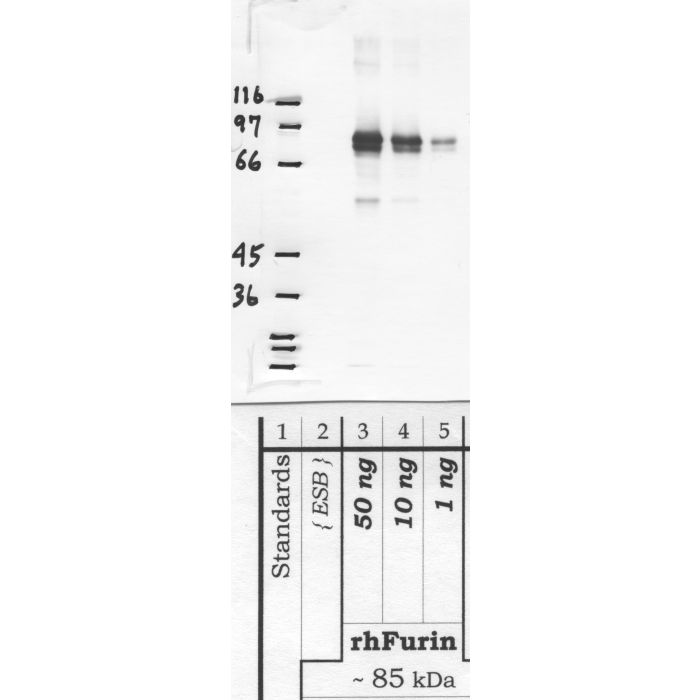
Western blot validation for RP-Furin — 2 panels across the domain-specific antibody variants. Each blot below shows the clone that validates a specific domain of the target protein.
Custom validation studies available on request — contact us.
Also known as:
- Furin
- FUR
- PACE
- Paired basic amino acid residue-cleaving enzyme
- Dibasic-processing enzyme
- PCSK3
- Proprotein convertase subtilisin/kexin type 3
- EC 3.4.21.75