
Anti-DPP-8 Rabbit Polyclonal Antibody
- Host
- Rabbit, Polyclonal
- Reactivity
- Validated- Human Potential-Mouse, Rat, Pan, Monkey, Dog
- UniProt
- Q6V1X1
- Size
- 100ug
- Cat. #
- RP3DPP8
In stock
- SKU
- RP-DPP8
Target Overview
Dipeptidyl peptidase 8 (DPP-8, EC 3.4.14.5; UniProt Q6V1X1) is a cytoplasmic serine protease that cleaves N-terminal dipeptides from substrates with proline or alanine at the P1' position. As a member of the S9B prolyl peptidase family, DPP-8 shares structural homology with DPP-4 and DPP-9 but lacks transmembrane domains and functions primarily as an intracellular regulator. Beyond its catalytic activity, DPP-8 serves a critical non-substrate role as a constitutive suppressor of NLRP1 and CARD8 inflammasomes in resting monocytes and macrophages. By sequestering the cleaved C-terminal fragments of NLRP1 and CARD8 in ternary complexes, DPP-8 prevents inflammasome oligomerization and caspase-1-dependent pyroptosis under basal conditions. Inhibition of DPP-8 triggers rapid pyroptotic cell death, a mechanism now exploited in cancer immunotherapy and studied in neurodegenerative disease models. The 898-amino-acid protein is expressed broadly across tissues, with particular relevance in immune cells where its regulatory function over inflammatory signaling is under active investigation.
Background
References
- Cui D et al (2026) MST1 promotes microglial pyroptosis and neuroinflammation in alzheimer's disease by regulating the novel DPP8/NLRP1/Caspase-1/GSDMD-N axis. J Neuroinflammation. PubMed · DOI
- Sewald L et al (2025) A Photocaged N-Phosphonopiperidinone as a Selective Photo-Cleavable DPP8/9 Inhibitor. Chembiochem. PubMed · DOI
- Filippi N et al (2025) Selective and Orally Bioavailable Dipeptidyl Peptidase 9 Inhibitors with Potent Pyroptosis Induction Properties. J Med Chem. PubMed · DOI
Additional Specifications
| Gene Symbol | DPP8 |
|---|---|
| UniProt ID | Q6V1X1 |
| Host Species | Rabbit |
| Species Reactivity | Validated- Human Potential-Mouse, Rat, Pan, Monkey, Dog |
| Pack Size | 100ug |
| Immunogen (Aminterminal end) | Immunogen is proprietary and confidential. Immunogen generated in amino acid region 1-51. |
| Immunogen (spacer region) | Immunogen is proprietary and confidential. Immunogen generated in amino acid region 420-530. |
| Immunogen (Catalytic domain) | Immunogen is proprietary and confidential. Immunogen generated in amino acid region 1-898. |
| Alternate Names | Dipeptidyl peptidase 8, DP8, DPP VIII, DPP8, Dipeptidyl peptidase IV-related protein 1, DPRP-1, Dipeptidyl peptidase VIII, Prolyl dipeptidase DPP8, EC 3.4.14.5 |
Frequently Asked Questions
What is the expected molecular weight for DPP-8 on Western blot?
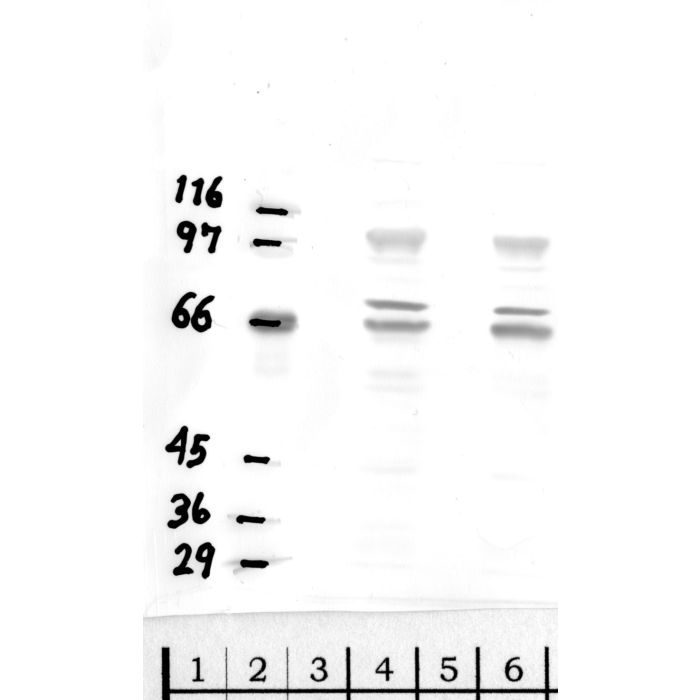
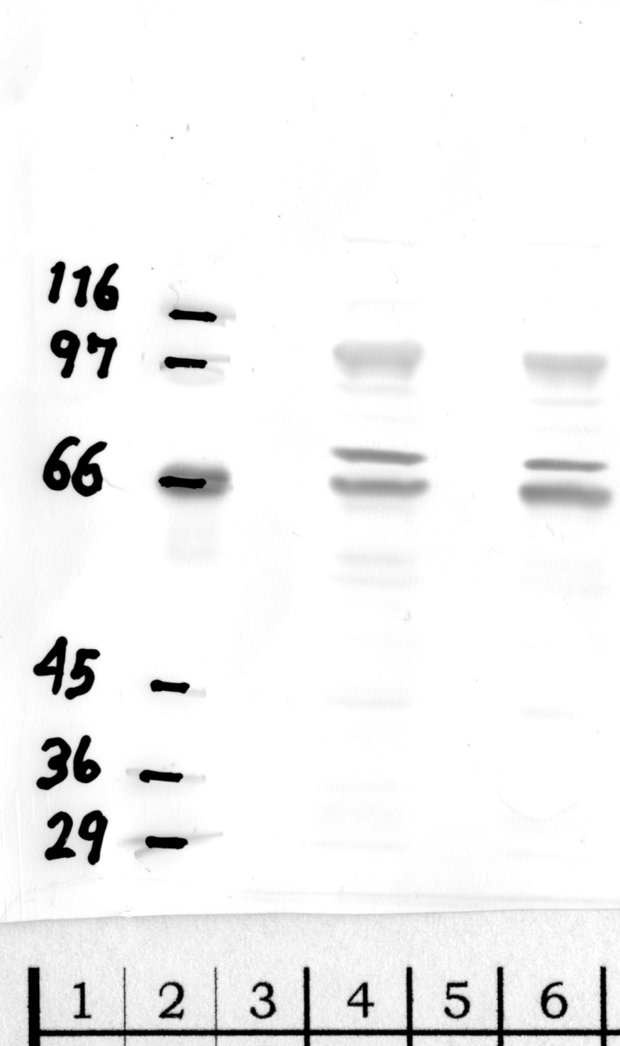
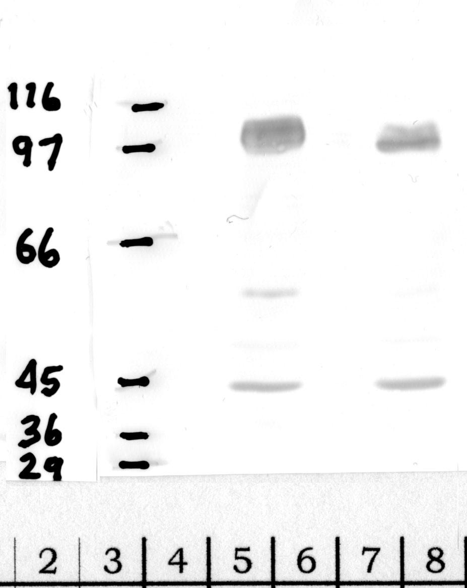
DPP-8 runs at approximately 103 kDa on SDS-PAGE, corresponding to its full-length 898-amino-acid sequence. You may observe a doublet or minor bands between 95–110 kDa depending on post-translational modifications or proteolytic processing in certain cell types. The antibody is raised against residues spanning a large portion of the protein, so it should detect full-length DPP-8 reliably. If you see lower bands around 50–60 kDa, consider the possibility of degradation during lysis; include protease inhibitors and keep samples cold throughout preparation.
Can this DPP-8 antibody distinguish between DPP-8 and DPP-9?
DPP-8 and DPP-9 share approximately 58% sequence identity and similar molecular weights, which can complicate specificity. This polyclonal was raised against a DPP-8-specific epitope region, and we have validated reactivity against human DPP-8 without cross-signal to recombinant DPP-9 in side-by-side Western blots. However, in complex lysates containing both enzymes, you should confirm identity by siRNA knockdown or include a DPP-9 antibody as a parallel control. DPP-4 is structurally related but membrane-bound and runs at 110 kDa, so it is less likely to cause confusion in cytosolic fractions.
What dilution should I start with for Western blot of DPP-8?
We recommend starting at 1:1000 in 5% non-fat milk or BSA in TBST for overnight incubation at 4°C. DPP-8 is expressed at moderate levels in most immortalized cell lines and primary monocytes, so this dilution balances signal and background. If your lysate is from a low-expressing primary tissue or you see weak signal, try 1:500. Conversely, if background is high in overexpression models, dilute to 1:2000. Always include a positive control lysate from THP-1 or HEK293 cells, which express endogenous DPP-8 robustly.
Is this antibody validated for mouse and rat DPP-8?
Human DPP-8 is validated by Western blot. Mouse and rat cross-reactivity are predicted based on high sequence homology in the immunogen region (greater than 85% identity), but we have not formally validated those species in-house. If you are working with mouse or rat samples, we suggest testing at 1:500 to 1:1000 and including a positive control from mouse RAW264.7 macrophages or rat spleen lysate. Report your results; cross-species performance can vary with post-translational modifications and fixation methods in IHC applications.
Can I use this DPP-8 antibody for immunofluorescence or IHC?
Triple Point Biologics antibodies are routinely validated for IHC and immunofluorescence in addition to Western blot. For DPP-8, start with a 1:100–1:200 dilution for IHC on paraffin-embedded sections after citrate-based antigen retrieval. In immunofluorescence, DPP-8 is cytoplasmic, so you should see diffuse staining throughout the cytosol in positive cells such as monocytes and macrophages. Avoid methanol fixation, which can mask epitopes; 4% paraformaldehyde for 10–15 minutes works well. Include a no-primary-antibody control to assess non-specific secondary binding.
What cell types or tissues have high endogenous DPP-8 expression?
DPP-8 is ubiquitously expressed but particularly abundant in immune cells. Human monocytes, macrophages, and dendritic cells show robust expression because DPP-8 constitutively suppresses NLRP1 and CARD8 inflammasomes. THP-1, U937, and primary peripheral blood mononuclear cells are reliable positive controls. Liver, kidney, and skeletal muscle also express moderate levels. In contrast, some epithelial lines and fibroblasts may show lower signal. If you are studying inflammasome regulation or pyroptosis, prioritize myeloid cell models where DPP-8 plays a well-characterized non-catalytic regulatory role.
How should I store this polyclonal DPP-8 antibody?
Store the antibody at –20°C in single-use aliquots to avoid repeated freeze-thaw cycles, which can reduce titer and increase aggregation. For short-term use over two to four weeks, you may keep a working aliquot at 4°C with 0.02–0.05% sodium azide as a preservative. Do not store diluted antibody in blocking buffer for more than one week; prepare fresh dilutions for each experiment when possible. If you observe precipitate after thawing, centrifuge briefly at 10,000 × g before use. Polyclonals are generally stable for at least two years under these conditions.
What controls should I include when probing for DPP-8 by Western blot?
Include a positive control lysate from cells known to express DPP-8, such as THP-1 monocytes or HEK293 cells. A negative control can be generated by siRNA knockdown of DPP8 in your cell line; residual signal after knockdown suggests off-target binding. If studying DPP-8 inhibition or inflammasome activation, compare untreated versus inhibitor-treated samples; DPP-8 protein levels should remain constant even when catalytic activity is blocked. Finally, run a no-primary control or isotype control to confirm that signal is antibody-dependent. These steps ensure that your band at 103 kDa is specific to DPP-8.
Western blot validation for RP-DPP8 — 3 panels across the domain-specific antibody variants. Each blot below shows the clone that validates a specific domain of the target protein.

Custom validation studies available on request — contact us.
Also known as:
- Dipeptidyl peptidase 8
- DP8
- DPP VIII
- DPP8
- Dipeptidyl peptidase IV-related protein 1
- DPRP-1
- Dipeptidyl peptidase VIII
- Prolyl dipeptidase DPP8
- EC 3.4.14.5