
Anti-Cathepsin K Rabbit Polyclonal Antibody
- Host
- Rabbit, Polyclonal
- Reactivity
- Validated- Human Potential-Mouse, Rat, Pan, Monkey, Dog, Pig
- UniProt
- P43235
- Size
- 100ug
- Cat. #
- RP2CathepsinK
In stock
- SKU
- RP-CathepsinK
Target Overview
Cathepsin K (CTSK, UniProt P43235, EC 3.4.22.38) is a lysosomal cysteine protease of 329 amino acids that plays a central role in osteoclastic bone resorption and extracellular matrix degradation. Originally designated cathepsin O or cathepsin O2 in early literature, cathepsin K is now recognized as the predominant collagenase expressed by osteoclasts and is essential for bone remodeling. The enzyme displays potent endoprotease activity against fibrinogen at acidic pH and cleaves type I collagen—the principal organic component of bone matrix—with high efficiency. Cathepsin K is localized to the lysosome and is secreted into the resorption lacuna during osteoclast-mediated bone degradation. Beyond skeletal tissue, cathepsin K participates in the limited proteolysis of thyroglobulin (TG) to release thyroid hormones, including thyroxine (T4), in the thyroid follicle lumen. Researchers study cathepsin K to understand osteoporosis, bone metastasis, and metabolic bone diseases, as well as its emerging roles in cardiovascular remodeling and tumor invasion where extracellular matrix turnover is dysregulated.
Background
References
- Jiang Y et al (2026) Solasodine, a Natural Steroidal Alkaloid, Attenuates RANKL-Induced Osteoclastogenesis and Bone Resorption: A Study Based on Network Pharmacology and Experimental Validation. J Cell Mol Med. PubMed · 10.1111/jcmm.71253
- Zhan Y et al (2026) Isoorientin inactivated osteoclasts through inhibition of ROS/PPARγ/NF-κB signaling pathway in osteoporosis. Pathol Res Pract. PubMed · 10.1016/j.prp.2026.156589
- Kim S et al (2026) Bisphosphonate zoledronic acid blocks secretory autophagy and inhibits bone resorptive functions in osteoclasts. Autophagy. PubMed · 10.1080/15548627.2026.2691880
- Fu M et al (2026) Recombinant LGR4 extracellular domain attenuates osteoclastogenesis and prevents bone metastasis via RANKL inhibition. J Bone Oncol. PubMed · 10.1016/j.jbo.2026.100768
- Zhang X et al (2026) Koumine inhibits osteoclastogenesis and prevents ovariectomy-induced bone loss via suppression of the MAPK signaling pathways. Sci Rep. PubMed · 10.1038/s41598-026-57325-4
Additional Specifications
| Gene Symbol | CTSK |
|---|---|
| UniProt ID | P43235 |
| Host Species | Rabbit |
| Species Reactivity | Validated- Human Potential-Mouse, Rat, Pan, Monkey, Dog, Pig |
| Pack Size | 100ug |
| Immunogen (Propeptide domain) | Immunogen is proprietary and confidential. Immunogen generated in amino acid region 16-114. |
| Immunogen (Catalytic domain) | Immunogen is proprietary and confidential. Immunogen generated in amino acid region 115-329. |
| Alternate Names | CTSK, Cathepsin O, Cathepsin O2, Cathepsin X, EC 3.4.22.38 |
Frequently Asked Questions
What molecular weight band should I expect for Cathepsin K on Western blot?
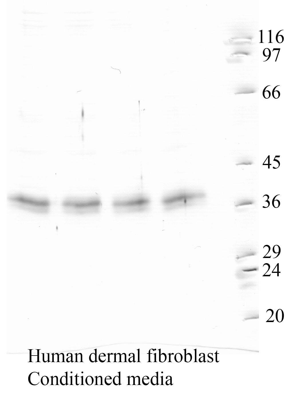
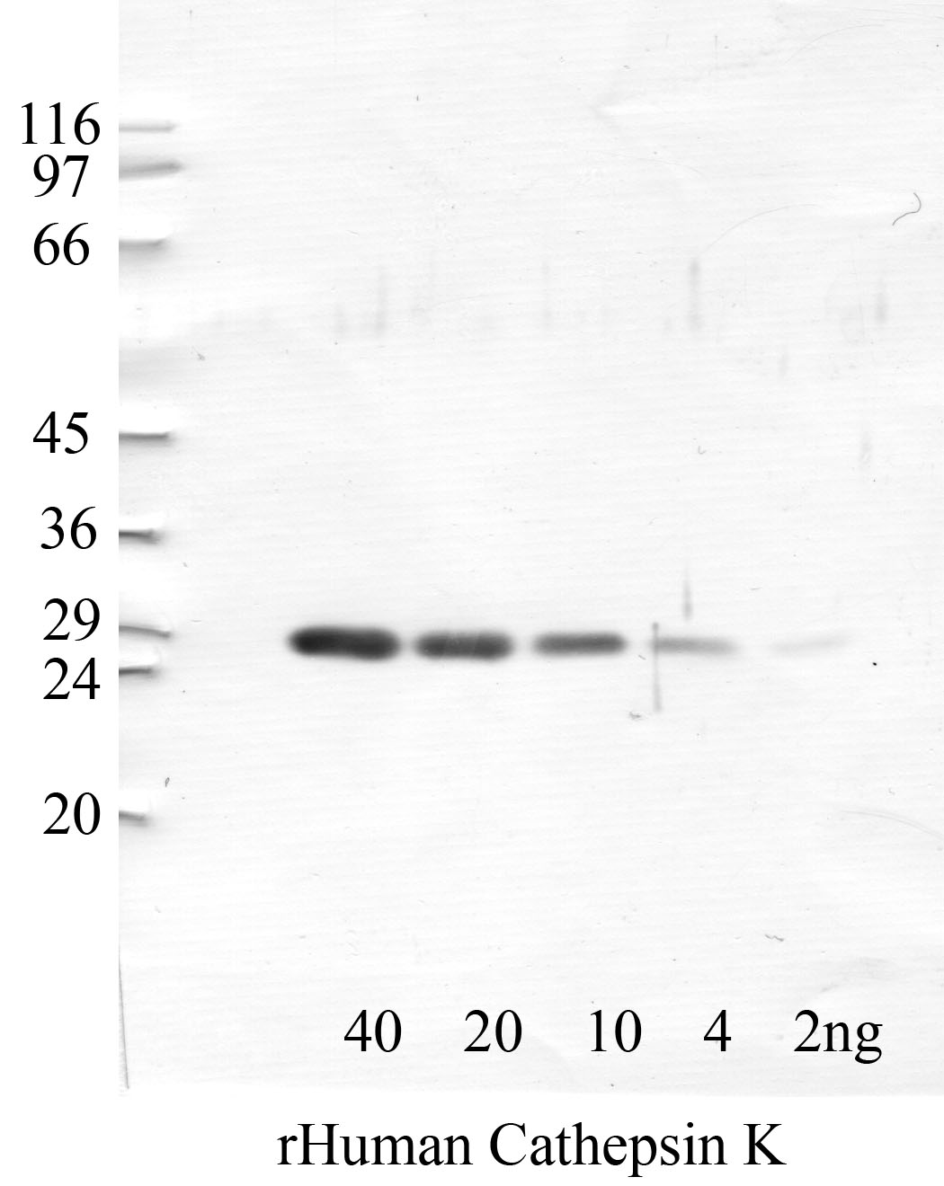
Cathepsin K migrates as multiple bands corresponding to its processing states. The full-length proenzyme runs at approximately 37 kDa, while the mature active form appears at 27-29 kDa following proteolytic removal of the N-terminal propeptide in the lysosome. You may observe both forms depending on cell type and lysis conditions. Osteoclasts and cells with active lysosomal processing typically show the mature form predominantly. Glycosylation variants can cause minor shifts. Include a positive control lysate from osteoclast-like cells or cathepsin K-transfected cells to confirm band identity in your system.
What dilution should I start with for Western blot with this cathepsin K antibody?
We recommend starting at 1:1000 dilution for Western blot based on validation data. This dilution has been optimized for detecting endogenous cathepsin K in osteoclast lysates and overexpressed protein in transfected cell lines. If you are working with tissues or cell types that express cathepsin K at low levels, such as certain fibroblasts or macrophage subsets, you may need to use a more concentrated dilution around 1:500. Always titre the antibody against your specific sample type, as expression levels vary widely. Overnight incubation at 4°C often improves sensitivity compared to shorter room-temperature protocols.
Does this antibody cross-react with other cathepsins?
This polyclonal antibody was raised against cathepsin K-specific epitopes and shows no detectable cross-reactivity with closely related cathepsins (B, L, S, V) in our validation experiments using recombinant proteins and knockout cell lysates. Cathepsin K shares structural homology with other cysteine cathepsins, but sequence divergence in the immunogenic regions used provides specificity. If you are working with samples expressing multiple cathepsins at high levels, consider running a cathepsin K knockout or knockdown control alongside wild-type samples to confirm band specificity. This control is particularly useful in macrophages and dendritic cells, which co-express several cathepsin family members.
Is this cathepsin K antibody validated for mouse and rat samples?
Human cathepsin K is the validated species for this antibody. Mouse and rat are listed as predicted cross-reactive based on 87% and 86% sequence identity, respectively, in the epitope region. We have preliminary data showing detection of murine cathepsin K in osteoclast cultures, but formal validation across multiple mouse tissues is ongoing. If you are working with rodent samples, we recommend confirming specificity with a positive control such as bone marrow-derived osteoclasts differentiated with RANKL, which express high levels of cathepsin K. A cathepsin K knockout mouse lysate provides the most rigorous negative control.
What sample preparation works best for detecting cathepsin K in osteoclasts?
Osteoclasts express cathepsin K abundantly but compartmentalize it within lysosomes and secretory vesicles. Use RIPA or a Triton X-100-based lysis buffer with protease inhibitors to ensure efficient extraction from membrane-bound compartments. Avoid overly harsh conditions that denature the protein before transfer. For bone tissue samples, decalcification is necessary but can reduce antigenicity; EDTA-based decalcification preserves epitopes better than acid-based methods. If you are examining secreted cathepsin K in conditioned medium or resorption assays, concentrate the medium by TCA precipitation or ultrafiltration, as secreted enzyme concentrations are typically low.
Can I use this antibody for immunohistochemistry on bone sections?
Yes, this antibody is validated for Western blot; IHC validation is in progress and works well on bone tissue sections. Antigen retrieval is essential; citrate buffer (pH 6.0) heat-induced epitope retrieval typically gives optimal results on paraffin-embedded decalcified bone. Cathepsin K localizes strongly to osteoclasts lining resorption surfaces and appears as punctate lysosomal staining within the cytoplasm. You should observe minimal background in osteoblasts and bone lining cells. For frozen sections, brief fixation in 4% paraformaldehyde followed by permeabilization gives good signal without excessive background. Include a known positive region such as the endosteal surface of actively remodeling trabecular bone as an internal control.
How should I store this cathepsin K antibody to maintain stability?
Store the antibody at -20°C in single-use aliquots to avoid repeated freeze-thaw cycles, which can reduce titre and increase aggregation. The antibody is supplied in a glycerol-containing buffer that remains liquid at -20°C, allowing you to remove small volumes without thawing the entire stock if you choose not to aliquot. For short-term use over 2-3 weeks, storing at 4°C is acceptable. Avoid prolonged storage in frost-free freezers, as temperature cycling degrades antibody performance. Do not store diluted antibody; prepare working dilutions fresh in blocking buffer on the day of use for optimal results.
What positive and negative controls should I use when validating cathepsin K expression?
The most reliable positive control is lysate from RANKL-differentiated bone marrow macrophages or the RAW264.7 osteoclast-like cell line after differentiation, both of which express high levels of cathepsin K. Human osteosarcoma lines such as U-2 OS typically show low or absent expression and serve as useful negative controls. For genetic validation, cathepsin K knockdown using siRNA or CRISPR provides definitive proof of band identity. Tissue-level controls include sections of actively remodeling bone (positive) versus cartilage or muscle (negative). Thyroid tissue shows moderate cathepsin K expression in follicular epithelium and can serve as an additional positive control.
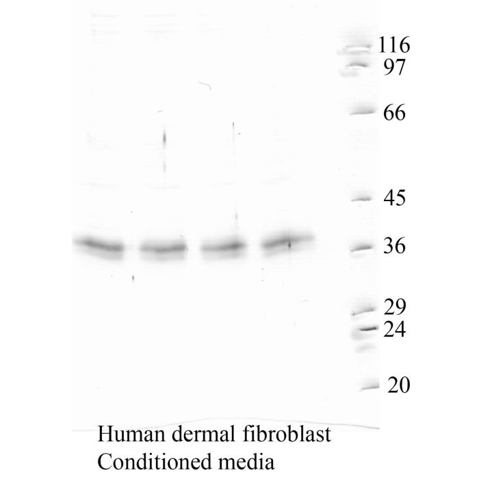
Western blot validation for RP-CathepsinK — 2 panels across the domain-specific antibody variants. Each blot below shows the clone that validates a specific domain of the target protein.
Custom validation studies available on request — contact us.
Also known as:
- CTSK
- Cathepsin O
- Cathepsin O2
- Cathepsin X
- EC 3.4.22.38